こんにちは!
運動器専門のリハビリスタッフです!!
いつもお世話になります。
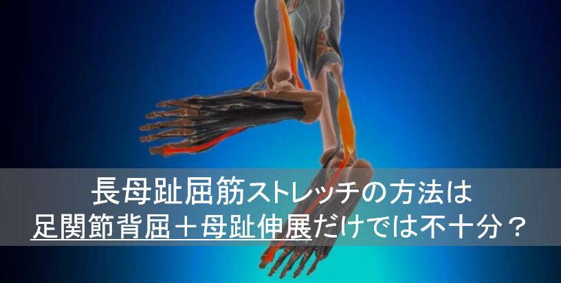
今回は、『長母趾屈筋ストレッチの方法は足関節背屈+母趾伸展だけでは不十分?』
について解説させていただきます。
長母趾屈筋(FHL)は距骨の後方を走行しており、距骨後方への移動を妨げます。
足関節背屈には距骨の後方移動を伴いますので、足関節背屈制限因子に長母趾屈筋はとても関連しています。
◆長母趾屈筋
起始:腓骨後面の下部2/3、下腿骨間膜の腓骨側
停止:母趾の末節骨底
作用:距腿関節の底屈、内反(回外)、母趾の中足趾節関節と趾節間(IP)関節の底屈、
内側縦足由弓の保持
神経支配:腓骨神経(L5-S2)
下記の論文で長母趾屈筋(FHL)の興味深い内容が報告されています。
![[商品価格に関しましては、リンクが作成された時点と現時点で情報が変更されている場合がございます。]](https://hbb.afl.rakuten.co.jp/hgb/1f7a7439.6deab8a7.1f7a743a.315603be/?me_id=1213310&item_id=21195057&pc=https%3A%2F%2Fthumbnail.image.rakuten.co.jp%2F%400_mall%2Fbook%2Fcabinet%2F4386%2F9784260054386_1_4.jpg%3F_ex%3D240x240&s=240x240&t=picttext "[商品価格に関しましては、リンクが作成された時点と現時点で情報が変更されている場合がございます。]")

Anatomical study of toe flexion by flexor hallucis longus
Mutsuaki Edama 1, Masayoshi Kubo 2, Hideaki Onishi 2, Tomoya Takabayashi 2, Erika Yokoyama 2, Takuma Inai 3, Hiroshi Watanabe 4, Satoshi Nashimoto 5, Ikuo Kageyama 6
Ann Anot 2016 Mar;204:80-5.
Abstract
Because connections exist between the flexor hallucis longus (FHL) and flexor digitorum longus (FDL), the FHL is surmised to exert a flexion action on the lesser toes, but this has not been studied quantitatively. The objectives of this study have thus been to clarify the types of FHL and FDL connections and branching, and to deduce the toe flexion actions of the FHL. One hundred legs from 55 cadavers were used for the study, with FHLs and FDLs harvested from the plantar aspect of the foot, and connections and branches classified. Image-analysis software was then used to analyze cross-sectional areas (CSAs) of each tendon, and the proportion of FHL was calculated in relation to flexor tendons of each toe. Type I (single slip from FHL to FDL tendon) was seen in 86 legs (86%), Type II (crossed connection) in 3 legs (3%), and Type III (single slip from FDL to FHL tendon) or Type IV (no connection between muscles) in 0 legs (0%). In addition, Type V (double slip from FHL to FDL tendon) was seen in 11 legs (11%), representing a new type not recorded in previous classifications. In terms of the various flexor tendons, the proportion of FHL showing tendons to toes 2 and 3 was high, at approximately 50-70%. Consequently, considering the branching type and proportion of CSA, the FHL was conjectured to not only act to flex the hallux, but also play a significant role in the flexion of toes 2 and 3. These results offer useful information for future clarification of the functional roles of tendinous slips from the FHL.
長母趾屈筋による足指屈曲の解剖学的研究
長母趾屈筋(FHL)と長趾屈筋(FDL)の間には接続があるため,FHLは足趾の屈曲作用を及ぼすと推測されるが,定量的な検討はなされていない。そこで,本研究では,FHLとFDLの接続と分岐のタイプを明らかにし,FHLの足指の屈曲作用を推測することを目的とした。研究には55人の献体から100本の脚を使用し,足底面からFHLとFDLを採取して,接続と分岐を分類した。その後,画像解析ソフトを用いて各腱の断面積(CSA)を解析し,各足指の屈筋腱に対するFHLの割合を算出した。その結果,Type I(FHLからFDLへの単一のすべり)が86脚(86%),Type II(交差した連結)が3脚(3%),Type III(FDLからFHLへの単一のすべり)またはType IV(筋肉間の連結なし)が0脚(0%)であった。さらに、タイプV(FHLからFDLへの腱の二重すべり)が11脚(11%)に見られ、これまでの分類では記録されていなかった新しいタイプであった。各屈筋腱については、FHLが第2趾、第3趾への腱を示す割合が高く、約50〜70%であった。その結果、分岐型やCSAの割合を考慮すると、FHLは母趾の屈曲に作用するだけでなく、第2趾、第3趾の屈曲にも重要な役割を果たしていると推測された。これらの結果は、今後、FHLからの腱索の機能的役割を明らかにする上で有用な情報となる。
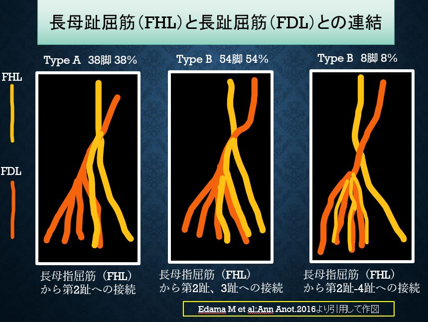
この論文の内容を一部引用し作図しました。
タイプAは長母指屈筋(FHL)から第2趾への連結:38脚、38%
タイプBはFHLから第2、第3趾への連結:54脚、54%
タイプCはFHLから第2-4趾への連結:8脚、8%
◆長趾屈筋
起始:腓骨後面の中央1/3
停止:第2-5末節骨底
作用:距腿関節の底屈、内反(回外)、第2-5趾の中足趾節(MP)関節・近位趾節間(PIP)
関節・遠位趾節間(DIP)の屈曲、
神経支配:腓骨神経(L5-S2)
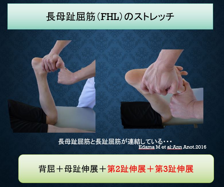
長母足屈筋(FHL)が第2趾、第3趾への腱を示す割合が、約50〜70%ということから、
長母趾屈筋のストレッチは足関節背屈+母趾伸展+【第2趾と第3趾伸展】がより伸張することが予測されます。
このように最新の解剖学を知識として得ると、技術がひとつ得ることができます。
今回は、『長母趾屈筋ストレッチの方法は足関節背屈+母趾伸展だけでは不十分?』
について解説させていただきました。